
Genetics
Progress in science depends on new techniques, new discoveries and new ideas, probably in that order.
— Sydney Brenner

Mus musculus


Mouse genetics tools in brain vasculature system field
- NG2DsRedBAC: NG2DsRedBAC transgenic mice is suitable for pericyte and Artery SMC labeling in central nervous system. NG2DsRedBAC transgenic mice express an optimized red fluorescent protein variant (DsRed.T1) under the control of the mouse NG2 (Cspg4) promoter/enhancer and may be useful for fluorescent labeling of NG2 cells (oligodendrocyte progenitor cells) in the central nervous system and NG2-expressing cells in other organs, as well as for isolating NG2 cell populations via FACS. [JAX Strain #:008241 | Genotyping Protocol] | DOI Link]
- Tek-Cre: The Tie2-Cre (Tek-Cre) transgene has the mouse endothelial-specific receptor tyrosine kinase (Tek or Tie2) promoter directing expression of Cre recombinase. These Tek-Cre transgenic mice are a Cre-lox tool useful for deletion of floxed sequences in endothelial cells during embryogenesis and adulthood. Cre recombinase activity is also detected in microglia in central nervous system. [JAX Strain #:008863 | Genotyping Protocol | DOI Link]
- Cdh5-PAC-CreERT2: [Genotyping Protocol | DOI Link]
- Pdgfrb-Cre: Pericyte labeling. DOI Link
- Pdgfrb-CreERT2: Pdgfrb-CreERT2 transgenic mice is suitable for Pericyte labeling, the result of Pdgfrb-CreERT2::Ai14 is very clean and specific. [JAX Strain #:029684 | Genotyping Protocol | DOI Link]
- BMX-CreERT2: Artery EC labeling. [Genotyping Protocol]
- Slco1c1-KI-P2A-iCreERT2: Brain specific EC labeling (including spinal cord). [Woo-Ping Ge lab, Design and characterized by Jun-Liszt Li, Contact | PMID: 33563078 | DOI Link]
Astrocyte labeling
-
Aldh1l1-EGFP: In this transgenic strain, Aldh1l1 promoter-active cells in the brain and spinal cord (the majority being astrocytes) express EGFP. Cre-mediated excision of the floxed EGFP/Stop enables DTA-mediated ablation of these cells. [JAX Strain #:026033 | Genotyping Protocol | PDF | PMID: 22745251 | DOI Link]
-
Aldh1l1-CreERT2: Aldh1l1-Cre/ERT2 BAC transgenic mice have tamoxifen-inducible Cre recombinase expression directed at high levels to the vast majority of astrocytes, with no detectable expression in neurons. These mice allow pan-astrocytic, specific and inducible genetic manipulations in vivo for studying astrocyte biology at different developmental stages in neural circuitry/synapses, behavior, disease and injury/trauma. [JAX Strain #:029655 | Genotyping Protocol]
-
mGFAP-Cre
-
hGfap-CreERT2:
- Park, Yongmin Mason, et al. “Astrocyte specificity and coverage of hGFAP-CreERT2 [Tg (GFAP-Cre/ERT2) 13Kdmc] mouse line in various brain regions.” Experimental Neurobiology 27.6 (2018): 508. [PMID: 30636902 | DOI Link]
-
Glast-CreERT2
-
Fgfr3-iCreERT2
Neuron labeling
- Thy1-YFP-M: These Thy1-GFP-M transgenic mice may be useful in neurobiological studies for fluorescent labeling of neural tissues, especially for mossy fibers in the cerebellum and intense, yet sparse, labeling of a variety of neuronal subsets. [JAX Strain #:007788 | Genotyping Protocol]
- Thy1-YFP-16J: These thy1-YFP-16 transgenic mice express yellow fluorescent protein at high levels in motor and sensory neurons, as well as in subsets of central neurons. This line provides a strong and specific vital marker for axons, and expression is strong from a mid-gestational stage into adulthood. [JAX Strain #:003709 | Genotyping Protocol]
- Gad1-EGFP: Also called Gad67-EGFP, Inhibitory GABAnergic neuron labeling. [JAX Strain #:007677 | Genotyping Protocol | DOI Link]
- Slc32a1-IRES2-FlpO: Slc32a1-IRES2-FlpO-D knock-in mice are designed to have optimized FLP recombinase expression directed to VGAT-expressing cells. FlpO expression/activity largely recapitulates the endogenous Slc32a1 gene in the brain - scattered Cre expression is observed throughout many brain regions, including the cortex, hippocampus, striatum and thalamus. These mice may be used to generate conditional mutations for studying gain-or-loss of function and/or fate mapping related to GABAergic neuronal function. [JAX Strain #:031331 | Genotyping Protocol | PMID: 30007418 | DOI Link]
- Vip-Cre: Vip+ Interneuron labeling.
- Sst-Cre: Sst+ Interneuron labeling.
- Som-Cre: Som+ Interneuron labeling.
- PV-Cre: PV+ Interneuron labeling.
- CamKIIa-Cre: Random insertion, Expression pattern in brain(Throughout the cortex, hippocampus, striatum, and other structures) | [Susumu Tonegawa] |
- Snap25-IRES2-Cre-D: Snap25-IRES2-Cre-D knockin mice have widespread Cre recombinase expression directed throughout the brain, without disrupting endogenous synaptosomal-associated protein 25 expression. These mice may be useful for studying t-SNARE proteins and synaptic vesicle/plasma membrane fusion. [Strain #:023525 | Genotyping Protocol | DOI Link]
- Snap25-IRES2-cre is superior to Syn1-cre as a pan-neuronal Cre driver.
- Slc17a7-IRES2-Cre: Slc17a7 (VGlut1), Excitatory neuron labeling [PMID: 25071457 | DOI Link]
- Drd1-Cre: These mice express Cre recombinase under the control of the mouse Drd1a (dopamine receptor D1A) promoter. DRD1A is s a G-protein coupled receptor involved in the regulation of neuronal growth and development. The D1 subtype is the most abundant dopamine receptor in the central nervous system. Homozygotes are viable and fertile. [MMRRC Strain #037156-JAX | DOI Link]
- Drd2-Cre
Microglia labeling
- CX3CR1-EGFP: [JAX Strain #:005582 | Genotyping Protocol]
- CX3CR1-iCreERT2: [JAX Strain #:021160 | Genotyping Protocol]
- Tmem119-P2A-iCreERT2: [JAX Strain #:031820 | Genotyping Protocol | DOI Link]
Oligodendrocytes
-
Oligo2-Cre: Oligo2-cre mice have a Cre recombinase inserted into the only exon of the oligodendrocyte transcription factor 2 Olig2 gene. The presence of cre abolishes expression of Olig2. OLIG2 is a basic helix-loop-helix transcription factor required for oligodendrocyte and motor neuron specification in the spinal cord, and for the development of somatic motor neurons in the hindbrain. Heterozygous mice are viable and fertile. [Strain #:025567 | Genotyping Protocol | DOI Link]
-
Plp1-CreERT2: These transgenic mice have a tamoxifen inducible Cre-mediated recombination system driven by the mouse Plp1, proteolipid protein (myelin) 1 promoter. When crossed with a strain containing a loxP site flanked sequence of interest, the offspring are useful for generating tamoxifen-induced, Cre-mediated targeted deletions. Tamoxifen administration allows for ablation of predetermined genes in oligodendrocytes and Schwann cells. [JAX Strain #:005975 | Genotyping Protocol | DOI Link]
Precusor cells
- Nestin-Cre: This strain is commonly known as nestin-Cre. Nestin promoter driven Cre recombinase is expressed in the central and peripheral nervous system, including neuronal and glial cell precursors. There are reports of Cre recombinase activity in a few isolated kidney and heart cells. Hemizygous nestin-cre mice exhibit mild hypopituitarism, growth retardation, impaired fear response, and a metabolic phenotype. Mice also show a strong impairment in the acquisition of both contextual- and cued-fear conditioning. [Strain #:003771 | Genotyping Protocol | DOI Link]
Reporter Lines
- Ai14(Rosa26-CAG-LSL-tdTomato-WPRE-pA)
- Ai47(Rosa26-CAG-LSL-emerald-TagGFP-hrGFP-WPRE-pA): The Rosa-CAG targeting vector was designed with (from 5’ to 3’) a CMV-IE enhancer/chicken beta-actin/rabbit beta-globin hybrid promoter (CAG), an FRT site, a loxP-flanked STOP cassette (with stop codons in all 3 reading frames and a triple polyA signal), triple GFP reporter genes (emerald, TagGFP (Aequorea macrodactyla GFP), and hrGFP (Humanized Renilla reniformis GFP)), a woodchuck hepatitis virus post-transcriptional regulatory element (WPRE; to enhance the mRNA transcript stability), a polyA signal, and an attB/attP-flanked PGK-FRT-Neo-polyA cassette. This entire construct was inserted between exons 1 and 2 of the Gt(ROSA)26Sor locus. [MGI:5750793 | PMID: 30007418 | DOI Link]
- Ai140(TIT2L-EGFP-ICL-tTA2): Ai140 (also called Ai140(TIT2L-EGFP-ICL-tTA2)) is a Cre-dependent, Tet-controllable fluorescent reporter line, created by targeted insertion at the Igs7 locus (TIGRE). Exposure to Cre recombinase removes both STOP cassettes - resulting in tTA2 expression and robust EGFP fluorescence. EGFP expression may be expected to be diminished/eliminated by doxycycline. [JAX Strain #:034100 | Genotyping Protocol | PMID: 30007418 | DOI Link]
Muscle cells
- HSA-Cre-ERT2: HSA-Cre-ERT2 mice (also called ACTA1-Cre-ERT2) express a tamoxifen-inducible cre/ERT2 fusion under the control of the human skeletal muscle α-actin (ACTA1) promoter and regulatory elements. Cre recombinase activity is observed only in skeletal muscles. This strain allows temporally controlled deletion of floxed genes in skeletal muscles. [Strain #:003771 | Genotyping Protocol | DOI Link]
Transgenic mouse line for disease modeling
-
5xFAD mice (Alzheimer disease model): These 5XFAD transgenic mice overexpress mutant human amyloid beta (A4) precursor protein 695 (APP) with the Swedish (K670N, M671L), Florida (I716V), and London (V717I) Familial Alzheimer’s Disease (FAD) mutations along with human presenilin 1 (PS1) harboring two FAD mutations, M146L and L286V. Both transgenes are regulated by the mouse Thy1 promoter to drive overexpression in the brain. 5XFAD mice recapitulate major features of Alzheimer’s Disease amyloid pathology and may be a useful model of intraneuronal Abeta-42 induced neurodegeneration and amyloid plaque formation. [JAX Strain #:006554 | Genotyping Protocol | PMID: 17021169 | DOI Link]
-
Braf-CA mice (for modeling bAVM disease):
- Li, Jun-Liszt, et al. “rAAV-miniBEND: A targeted vector for brain endothelial cell gene delivery and cerebrovascular malformation modeling.” bioRxiv (2025): 2025-06. [JAX Strain #:017837]
-
Rosa26-LSL-KRAS-G12D
Transgenic line for Calcium imaging
- Thy1-GCaMP6f: These transgenic mice, made by the Genetically-Encoded Neuronal Indicator and Effector (GENIE) Project, express the green fluorescent calcium indicator, GCaMP6f, in subsets of excitatory neurons in the brain and possibly other cell types and tissues. The GCaMP6f indicator is an ultrasensitive detector of single neuronal action potentials and has fast response kinetics. [JAX Strain #:024339 | Genotyping Protocol | PMID: 23868258 | DOI Link]
- Thy1-JRGECO1a
- Ai96(Rosa26-CAG-LSL-GCaMP6s)
Transgenic line for genome editing
- Rosa26-CAG-LSL-SpCas9-P2A-EGFP(Rosa26-floxed STOP-Cas9 knockin): These CRISPR/Cas9 knockin mice have Cre recombinase-dependent expression of CRISPR associated protein 9 (cas9) endonuclease, a 3X-FLAG epitope tag and EGFP directed by a CAG promoter. Expression of Cas9 and EGFP is prevented by an upstream Lox-Stop-Lox (LSL) sequence. When used in combination with single guide RNAs and a Cre source, they allow editing of single or multiple mouse genes in vivo or ex vivo. [Strain #:024857 | Genotyping Protocol | PMID: 25263330 | DOI Link]
- Rosa26-CAG-SpCas9-P2A-EGFP(Rosa26-Cas9 knockin): These CRISPR/Cas9 knockin mice constitutively express CRISPR associated protein 9 (cas9) endonuclease, a 3X-FLAG epitope tag and EGFP in a widespread fashion under the direction of a CAG promoter. Breeding to a germ-line expressed Cre strain during development removed a Lox-Stop-Lox (LSL) sequence to allow ubiquitous expression of the knockin construct. When used in combination with single guide RNAs, they allow editing of single or multiple mouse genes in vivo or ex vivo. [Strain #:024858 | Genotyping Protocol | PMID: 25263330 | DOI Link]
Ref. public websites
- The Jackson Laborary: The laboratory is also the world’s source for more than 8,000 strains of genetically defined mice, and serves as a worldwide center for scientific courses, conferences, training, and education.
- Mouse Genome Informatics: MGI is the international database resource for the laboratory mouse, providing integrated genetic, genomic, and biological data to facilitate the study of human health and disease.
- Allen Institute for Brain Research
Ref. Labs
- Susumu Tonegawa Lab, MIT
- Joe Z. Tsien Lab, Princeton University
- Subregion- and Cell Type–Restricted Gene Knockout in Mouse Brain with phage P1–derived Cre/loxP recombination system
- Tsien, Joe Z., et al. “Subregion-and cell type–restricted gene knockout in mouse brain.” Cell 87.7 (1996): 1317-1326. [PDF | PMID: 8980237 | DOI Link]
- Masashi Yanagisawa Lab, The International Institute for Integrative Sleep(IIIS)
- Z. Josh Huang Lab, Duke University
Ref. Protocols
1. Long ssDNA for Knockin
- Guide-it Long ssDNA Production System v2 [Takara Cat. #: 632666 | User Manual]
- The kit employs a simple and fast method that involves conversion of a dsDNA PCR product into ssDNA via selective digestion of either the sense or the antisense strand. Following digestion, ssDNA products are purified using silica membrane spin columns included with the kit.
- It has been demonstrated that ssDNA offers two key advantages over dsDNA templates for precise genome editing applications: greatly reduced toxicity and a much lower likelihood of random or off-target integration.
- Easi-CRISPR
- Miura, Hiromi, et al. “Easi-CRISPR for creating knock-in and conditional knockout mouse models using long ssDNA donors.” Nature protocols 13.1 (2018): 195-215. [PDF | PMID: 29266098 | DOI Link]
2. Genetic screen
-
ENU mutagenesis, forward genetic screen
- Funato H, Miyoshi C, Fujiyama T, et al. Forward-genetics analysis of sleep in randomly mutagenized mice[J]. Nature, 2016, 539(7629): 378-383. [PDF | PMID: 27806374 | DOI Link]
-
CRISPR based pool screen, in cell line.
Vendor company
Rat
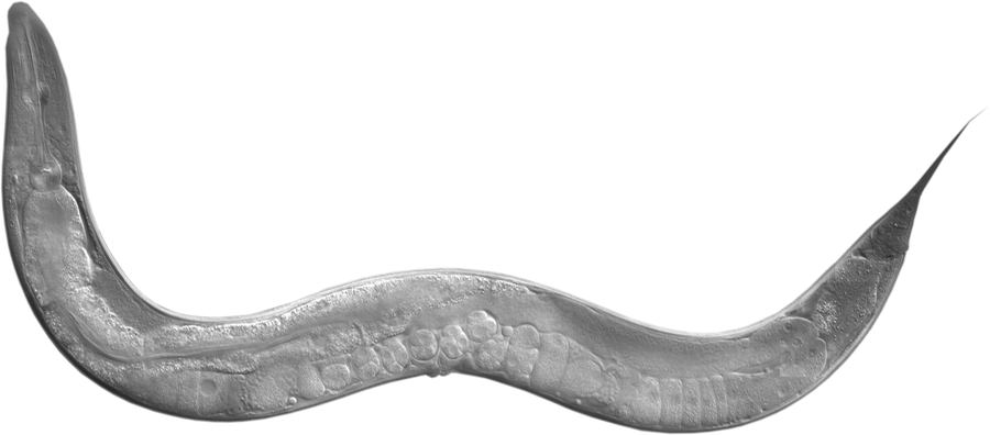
Caenorhabditis elegans (C. elegans)
Pioneers
-
Sydney Brenner (1927-2019), The Molecular Sciences Institute, Berkeley, CA, USA
-
Victor Ambros (1953- ), UMass Chan Medical School, Worcester, MA, USA
- [Biography]: Victor Ambros grew up in Vermont and graduated from MIT in 1975. He did his graduate research (1976-1979) with David Baltimore at MIT, studying poliovirus genome structure and replication. He began to study the genetic pathways controlling developmental timing in the nematode C. elegans as a postdoc in H. Robert Horvitz’s lab at MIT, and continued those studies while on the faculty of Harvard (1984-1992), Dartmouth (1992-2007), and the University of Massachusetts Medical School (2008-present). In 1993, members of the Ambros lab identified the first microRNA, the product of lin-4, a heterochronic gene of C. elegans. Since then, the role of microRNAs in development has been a major focus of his research.
- Lab website
Drosophila melanogaster
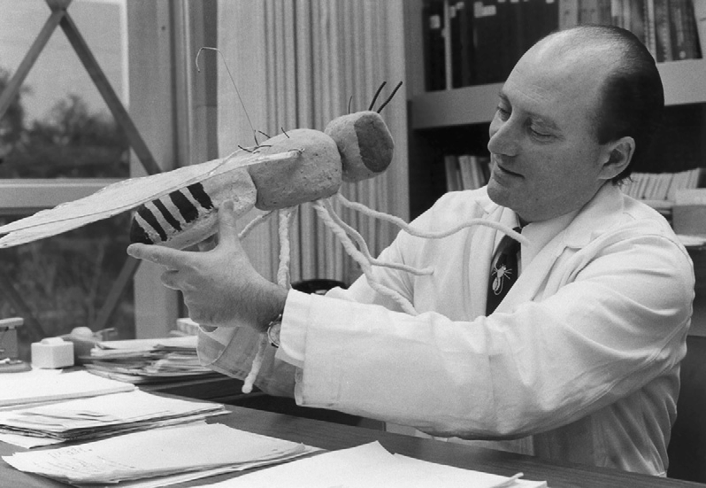 Seymour Benzer with mega-Drosophila, 1974.
Pioneers
-
Seymour Benzer (1921-2007), California Institute of Technology
-
Norbert Perrimon, Harvard Medical School/HHMI [Lab Link]
- Developed the Gal4/UAS system, with Andrea Brand
– Brand A H, Perrimon N. Targeted gene expression as a means ofaltering cell fates and generating dominant phenotypes. development, 1993,118(2): 401-415.[PMID:8223268 | DOI Link] - Developed the RNAi screen system in Drosophila cells
– Boutros M, Kiger A A, Armknecht S, et al. Genome-wide RNAi analysis of growth and viability in Drosophila cells. Science, 2004, 303(5659): 832-835. [PMID:14764878 | DOI Link]
- Developed the Gal4/UAS system, with Andrea Brand
Ref. Websites
- FlyBase: A Database of Drosophila Genes & Genomes
- DRSC/TRiP Functional Genomics Resources & DRSC-BTRR: Drosophila RNAi Screening Center (DRSC), Transgenic RNAi Project (TRiP) and Drosophila Research & Screening Center-Biomedical Technology Research Resource (DRSC-BTRR)
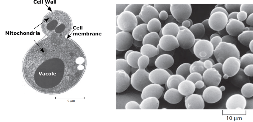
Yeast
Ref protocol
- EMS based Genetic screen
-
Tsukada, Miki, and Yoshinori Ohsumi. “Isolation and characterization of autophagy-defective mutants of Saccharomyces cerevisiae.” FEBS letters 333.1-2 (1993): 169-174. [PMID: 8224160 | DOI Link]
- Key details: BJ3505 cells were grown in YEPD to stationary phase, and then suspended in 340 μl of 0.1 M sodium phosphate buffer (pH 7.0) and treated with 10 μl EMS for 60 min at 30°C; the survival rate was 40-50%. The mutagenized culture (20 μl) was diluted with 780 μl of 5% sodium thiosulfate and spread on YEPD plates.
-
L.H. Hartwell,J. Culotti, & B. Reid, Genetic Control of the Cell-Division Cycle in Yeast, I. Detection of Mutants, Proc. Natl. Acad. Sci. U.S.A. 66 (2) 352-359, [DOI Link]
- Key details: Mutant isolation: A culture of S. cerevisiae, strain A364A, was grown from a small inoculum overnight at 36 ℃. in YM-1. While still growing logarithmically, 50 ml of culture was shifted to 23 ℃. and 0.2 ml of a solution containing 4 mg (per ml) of N-methyl-N’-nitro-N-nitrosoguanidine was added. Samples containing 0.5 ml of the culture were immediately distributed to a large number of tubes. The tubes were then rotated for a period of 5 hr (survival, 0.2 to 1.0%), after which time samples were removed, diluted, plated on YEPD-TAU plates, and incubated at 23 ℃. When colonies appeared, the pattern was replicated onto two plates, the first of which was incubated at 36 C and the second of which was incubated at 23 ℃. Colonies which grew up on the latter but not the former (approximately 1% of the total number of colonies) were picked, diluted with water, and streaked onto two YEPD-TAU plates, which were again incubated at the two temperatures. Clones which formed approximately 1,000 colonies on the 23 ℃. plate and no colonies on the 36 C plate were picked from the low-temperature plate and designated as ts- mutants. Only one or two mutants were isolated from a single mutagen-treated culture tube.
-
Ref. Websites
- YeastGFP: The YeastGFP database (the Yeast GFP Fusion Localization Database) of global analysis of protein localization studies in the budding yeast, S. cerevisiae, was originally designed and built by the laboratories of Erin O’Shea and Jonathan Weissman at the University of California, San Francisco. It is now hosted by SGD.
Zebrafish
Ref. Websites
- ZFIN: The Zebrafish Information Network (ZFIN) is the database of genetic and genomic data for the zebrafish (Danio rerio) as a model organism. ZFIN provides a wide array of expertly curated, organized and cross-referenced zebrafish research data.
Ref protocol
- Genetic screen
- ENU mutagenesis, forward genetic screen
- Patton E E, Zon L I. The art and design of genetic screens: zebrafish[J]. Nature Reviews Genetics, 2001, 2(12): 956-966. [PDF | PMID: 11733748 | DOI Link]
Nobel laureates

-
Victor Ambros, UMass Chan Medical School, Worcester, MA, USA
Nobel Prize in Physiology or Medicine 2024
A marvelous unfolding story of microRNAs [Nobel Lecture video | Read the Lecture | Source | Lab Link] -
Gary Ruvkun, Massachusetts General Hospital, Boston, MA, USA; Harvard Medical School, Boston, MA, USA
Nobel Prize in Physiology or Medicine 2024
A vast and ancient hidden world of microRNAs across the eukaryotes [Nobel Lecture video | Source | Lab Link] -
Michael W. Young, Rockefeller University, New York, NY, USA
The Nobel Prize in Physiology or Medicine 2017
Time Travels: A 40 Year Journey from Drosophila’s Clock Mutants to Human Circadian Disorders [Nobel Lecture video | Lecture Slides | Read the Lecture | Source] -
Jeffrey C. Hall, University of Maine, Maine, ME, USA Nobel Prize in Physiology or Medicine 2017
The Little Flies and their Genes, all the way to uncovering Mysteries of the Circadian Clock [Nobel Lecture video | Read the Lecture | Source] -
Michael Rosbash, Brandeis University, Waltham, MA, USA; Howard Hughes Medical Institute, USA Nobel Prize in Physiology or Medicine 2017
The Circadian Clock, Transcriptional Feedback and the Regulation of Gene Expression [Nobel Lecture video | Lecture Slides | Read the Lecture | Source] -
Yoshinori Ohsumi, Tokyo Institute of Technology, Tokyo, Japan
The Nobel Prize in Physiology or Medicine 2016
Molecular Mechanisms of Autophagy in Yeast [Nobel Lecture video | Lecture Slides | Read the Lecture | Source] -
Randy W. Schekman, University of California, Berkeley
The Nobel Prize in Physiology or Medicine 2013
Genes and Proteins That Control the Secretory Pathway [Nobel Lecture video | Lecture Slides | Read the Lecture | Source] -
Bruce A. Beutler, University of Texas Southwestern Medical Center at Dallas, Dallas, TX, USA; The Scripps Research Institute, La Jolla, CA, USA
Nobel Prize in Physiology or Medicine 2011
How Mammals Sense Infection: From Endotoxin to the Toll-like Receptors [Nobel Lecture video | Lecture Slides | Read the Lecture | Source] -
Robert G. Edwards (1925-2013), University of Cambridge, Cambridge, United Kingdom
The Nobel Prize in Physiology or Medicine 2010
for the development of in vitro fertilization [Nobel Lecture video | Lecture Slides | Read the Lecture | Source] -
Andrew Z. Fire, Stanford University School of Medicine, Stanford, CA, USA
The Nobel Prize in Physiology or Medicine 2006
Gene Silencing by Double Stranded RNA [Nobel Lecture video | Lecture Slides | Read the Lecture | Source] -
Craig C. Mello, University of Massachusetts Medical School, Worcester, MA, USA
Nobel Prize in Physiology or Medicine 2006
Return to the RNAi World: Rethinking Gene Expression and Evolution [Nobel Lecture video | Lecture Slides | Read the Lecture | Source | Lab Link] -
Leland H. Hartwell, Fred Hutchinson Cancer Research Center, Seattle, WA, USA
Nobel Prize in Physiology or Medicine 2004
Yeast and Cancer [Nobel Lecture video | Read the Lecture | Source] -
Sydney Brenner (1927-2019), The Molecular Sciences Institute, Berkeley, CA
The Nobel Prize in Physiology or Medicine 2002
Nature’s Gift to Science [Nobel Lecture video | Read the Lecture | Source] -
Barbara McClintock (1902-1992), Cold Spring Harbor Laboratory, Cold Spring Harbor, NY, USA
The Nobel Prize in Physiology or Medicine 1983
The Significance of Responses of the Genome to Challenge [Nobel Lecture video | Read the Lecture | Source] -
Max Delbrück (1906-1981), California Institute of Technology (Caltech), Pasadena, CA, USA
The Nobel Prize in Physiology or Medicine 1969
A Physicist’s Renewed Look at Biology–Twenty Years Later [Read the Lecture | Source] -
Alfred D. Hershey (1908-1997), Carnegie Institution of Washington, Long Island, New York, NY, USA
The Nobel Prize in Physiology or Medicine 1969
Idiosyncrasies of DNA Structure [Read the Lecture | Source] -
Salvador E. Luria (1912-1991), Massachusetts Institute of Technology (MIT), Cambridge, MA, USA
The Nobel Prize in Physiology or Medicine 1969
Phage, colicins and macroregulatory phenomena [Read the Lecture | Source] -
Thomas Hunt Morgan (1866-1945), California Institute of Technology (Caltech), Pasadena, CA, USA
The Nobel Prize in Physiology or Medicine 1933
The Relation of Genetics to Physiology and Medicine [Read the Lecture | Source]