Microscopic Imaging
Believing is seeing and seeing is believing.
— Tom Hanks

Confocal Microscopy
Commercial
Leica: Leica-SP8 (Archived Product Replaced by STELLARIS 5 & STELLARIS 8)
Carl Zeiss: LSM 880, with Airyscan: The Zeiss LSM880 with Ariyscan 2 is an inverted confocal microscope. The system is equipped with 7 laser lines 405nm, 458nm, 488nm, 514 nm, 561nm, 594nm and 633 nm. The newly designed excitation beam is elongated in y which acquires four lines of image information instead of only one with just one horizontal scanner movement. [Standard Operation Protocol | Detailed Configuration]
Information about Microscope Objectives
- Please refer to the following inventory for all of the objectives available at the Janelia imaging core facility. [Inventory list | Zeiss confocal objectives]
- Manufacturer brochures for objective types can also be viewed: Zeiss Objectives, Nikon Objectives, and Olympus Objectives.
SpectraViewers
- Fluorescence SpectraViewer, ThermoFisher [Manual]
- SearchLight, Semrock [Manual]
- Fluorescence Spectrum Viewer, BD Biosciences: The BD Spectrum Viewer is a tool that depicts the excitation and emission curves of fluorochromes common to flow cytometry. This tool can be used to determine appropriate filters to detect a fluorochrome as well as fluorochrome compatibility and fluorescent spillover.
Staining
- Fluorescence Dye and Filter Database, Zeiss
- Cell Staining Simulation Tool, Life Technologies
Two-Photon Microscopy
Protocol for mouse cranial window surgery, by Jun-Liszt Li [Download PDF]
Protocol for in vivo calcium imaging of neuronal populations, by Jun-Liszt Li [Download PDF]
Lab DIY
Thorlabs: https://www.thorlabs.com/index.cfm
Ti:Sapphire Femtosecond Laser: From Thorlabs
AO (Adaptive optics) in 2PFM and for neurobiology
Commercial
Olympus: FVMPE-RS
Leica: Leica-MP-4Tune
Scientifica: Multiphoton Imaging System
TMC: TMC combines state-of-the-art floor vibration cancellation technology with a commitment to manufacturing. Link
Ref labs: Ji Na lab, University of California, Berkeley
Janelia farm
Ref softwares:
- Suite2p [Offical website | Github]
- CalmAn [Online documentation | Github]
- CNMF_E [Github | DOI Link]
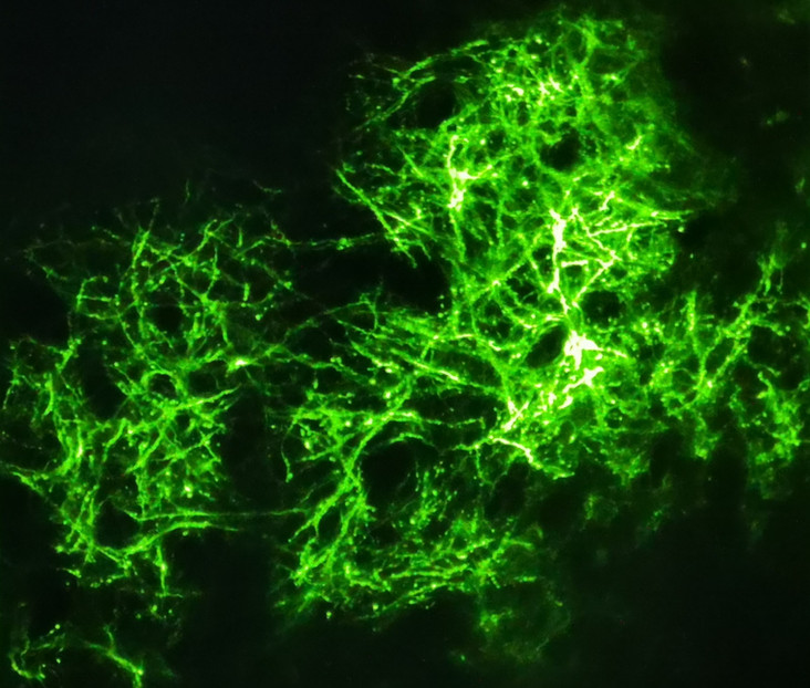
In this talk, two-photon microscopy-which uses strong pulsed infrared lasers to produce detailed images inside biological samples-is introduced. It can be utilized to image inside of living animals as well as thick tissue specimens.
Light-sheet Microscopy
Lab DIY
Thorlabs:
Tiling Light-Sheet Microscopy(TLSM): Liang Gao lab, Westlake University
Open-top Light-Sheet Microscopy: Jonathan T. C. Liu Lab, University of Washington
- Glaser, Adam K., et al. “A hybrid open-top light-sheet microscope for versatile multi-scale imaging of cleared tissues.” Nature Methods 19.5 (2022): 613-619. [PDF | Link]
Axial-Sweeping technology: Fiolka Reto lab,
- Dean, Kevin M., et al. “Deconvolution-free subcellular imaging with axially swept light sheet microscopy.” Biophysical journal 108.12 (2015): 2807-2815. [PDF | Link]
Mesoscale selective plane illumination microscopy(mesoSPIM): Official website
- Voigt, Fabian F., et al. “The mesoSPIM initiative: open-source light-sheet microscopes for imaging cleared tissue.” Nature methods 16.11 (2019): 1105-1108. [PDF | Link]
Commercial
Lavision BioTec Inc:
UltraMicroscope Light Sheet Microscope
3i Inc: Lattice Light Sheet Microscope
ZEISS: ZEISS Lattice Lightsheet 7
Ref labs: Philipp Keller lab, Janelia farm
Eric betzig lab, University of California, Berkeley
Reto Fiolka lab, UTSW
Liang Gao Lab, Westlake University
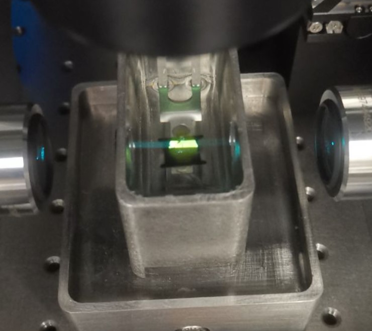
The innovative method of light sheet microscopy, also known as selective plane illumination, is discussed in this talk (SPIM). This allows for long-term 3D imaging of dense specimens like growing embryos with less photobleaching and phototoxicity because it uses two objectives-one to illuminate the sample and the other to image it.
Super-resolution microscopy
In comparison to traditional microscopy with diffraction-limited optics, a range of contemporary techniques are surveyed in this presentation. These include various patterns of illumination (such as STED and SIM microscopy) or methods that construct images by randomly turning on single fluorescent molecules and precisely localizing each molecule in space (STORM, PALM, FPALM).
During 20 years of friendship, Betzig and Hess collaborated to create the first super-high-resolution PALM microscope after working independently and jointly in academia and industry. They describe the journey to us, emphasizing how their diverse and unusual backgrounds gave them the necessary abilities to complete the project.
Ref labs: Xiaowei Zhuang Lab, Harvard University
Eric betzig lab, University of California, Berkeley
Liangyi Chen, PKU
Dong Li, IBP
Electron Microscopy
Ref labs:
- Thomas Reese, National Institutes of Health and John Heuser, Washington University in St. Louis
Thomas Reese studies synaptic structure and function using advanced light and electron microscopy techniques. John Heuser developed and spent his career using quick-freeze, deep-etch electron microscopy to study all aspects of cell biology.\
- Imaging Synaptic Vesicle Transmission: Two pioneering electron microscopists, John Heuser and Tom Reese, reminisce about their early attempts to image synaptic vesicle transmission. (iBiology interview, recorded in July 2015: [Part 1: Imaging synaptic vesicle transmission | Part 2: The Future of Electron Microscopy | Part 3: Why Collaborate? | Source])
Croy-Electron Microscopy(cryo-EM)
Ref labs:
- Yifan Cheng Lab, UCSF
Atomic structures of TRPV1: With David Julius’s laboratory at UCSF
[Liao, et al. 2016, Nature; Cao et al. 2013 Nature and Gao et al. 2016, Nature]
iBiology Talks: [Part 1: Single Particle Cryo-EM | Part 2: Single particle Cyro-EM of membrane proteins] - Niko Grigorieff Lab
- Grant Jensen Lab, Caltech
- Course: “Getting Started in Cryo-EM”
- Part 1: Currents, coils, knobs and names: Basic anatomy of the electron microscope [PDF]
- Part 2: Fourier transforms and reciprocal space for beginners [PDF]
- Part 3: Image formation [PDF]
- Part 4: Fundamental challenges in biological EM [PDF]
- Part 5: Tomography [PDF]
- Part 6: Single-particle analysis [PDF]
- Part 7: 2-D crystallography [PDF]
Ref softwares:
- MotionCorr: This program corrects whole frame image motion recordded with dose fractionated image stack. MotionCor2
- Xueming Li, Paul Mooney, Shawn Zheng, Chris Booth, Michael B. Braunfeld, Sander Gubbens, David A. Agard and Yifan Cheng (2013) Electron counting and beam-induced motion correction enables near atomic resolution single particle cryoEM. Nature Methods, 10, 584-590. PMID: 23644547
- GeFREALIGN: FREALIGN is a program developed by Niko Grigorieff laboratory for high-resolution refinement of 3D reconstruction from cryoEM of single particles.
- Xueming Li, Nikolaus Grigorieff and Yifan Cheng (2010) GPU-enabled FREALIGN: Accelerating single particle 3D reconstruction and refinement in Fourier space on graphics processors. Journal of Structural Biology, advanced publication online June 14 2010. PMID: 20558298
Ref database:
Open Source Softwares
- Fuji: Fuji is just imagej. Link
- Imaris Viewer: https://imaris.oxinst.com/imaris-viewer
Imaris 9.0 tutorial [PDF] - Cellfinder: https://github.com/SainsburyWellcomeCentre/cellfinder
- BrainRender: https://github.com/BrancoLab/BrainRender
- Allen Developing Brain atlas: https://developingmouse.brain-map.org/static/atlas
- NeuronStudio: A free platform that provides tools for 2D/3D visualization as well as manual and automatic detection/tracing of dendritic structures. Link
- yEd: A free, open-source, cross-platform graph editor used to quickly and effectively generate high-quality diagrams.
Nobel laureates
-
Jacques Dubochet, University of Lausanne, Lausanne, Switzerland
The Nobel Prize in Chemistry 2017
Early cryo-electron microscopy [Nobel Lecture video | Lecture Slides | Read the Lecture | Source] -
Joachim Frank, Columbia University, New York, NY, USA
The Nobel Prize in Chemistry 2017
Single-Particle Reconstruction – Story in a Sample [Nobel Lecture video | Lecture Slides | Read the Lecture | Source] -
Richard Henderson, MRC Laboratory of Molecular Biology, Cambridge, United Kingdom
The Nobel Prize in Chemistry 2017
From Electron Crystallography to Single Particle cryoEM [Nobel Lecture video | Lecture Slides | Read the Lecture | Source] -
Eric betzig, Janelia/University of California, Berkeley
The Nobel Prize in Chemistry 2014
Single Molecules, Cells, and Super-Resolution Optics [Nobel Lecture video | Lecture Slides | Read the Lecture | Source] -
William E. Moerner, Stanford University, Stanford, CA, USA
The Nobel Prize in Chemistry 2014
Single-Molecule Spectroscopy, Imaging, and Photocontrol: Foundations for Super-Resolution Microscopy [Nobel Lecture video | Lecture Slides | Read the Lecture | Source] -
Maria Goeppert Mayer, University of California, San Diego, CA, USA
The Nobel Prize in Physics 1963
The Shell Model
